
پەڕگە:Animal Cell.svg

Size of this PNG preview of this SVG file: ٨٠٠ لە ٤٦٢ پیکسەڵ. ڕێزەلووشنەکانی تر: ٣٢٠ لە ١٨٥ پیکسەڵ | ٦٤٠ لە ٣٦٩ پیکسەڵ | ١٬٠٢٤ لە ٥٩١ پیکسەڵ | ١٬٢٨٠ لە ٧٣٩ پیکسەڵ | ٢٬٥٦٠ لە ١٬٤٧٨ پیکسەڵ | ١٬٤٠٥ لە ٨١١ پیکسەڵ.
پەڕگەی سەرەکی (پەڕگەی SVG، بە ناو ١٬٤٠٥ × ٨١١ پیکسەڵ، قەبارەی پەڕگە: ٤٥٧ کیلۆبایت)
کورتە
| وەسف |
English: A reworked version of File:Biological_cell.svg.
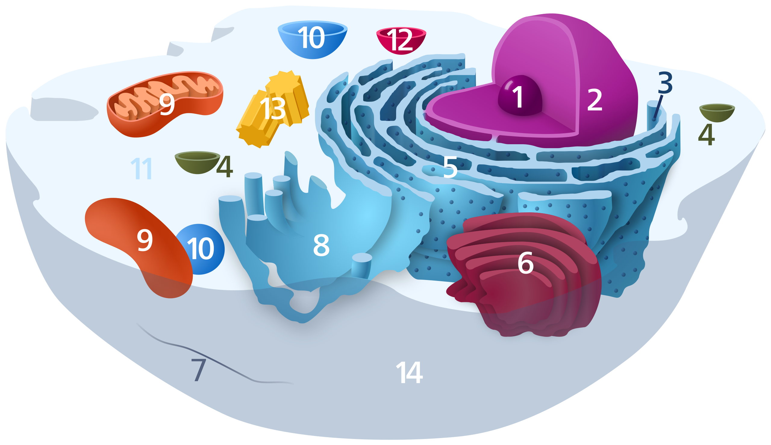
Diagram of a typical animal cell. Organelles are labelled as follows:
العربية: رسم تخطيطي للخلية الحيوانية
Català: Dibuix esquemàtic d'una cèl·lula animal típica:
Español: Diagrama de una célula animal típica:
ਪੰਜਾਬੀ: ਕਿਸੇ ਮਿਸਾਲੀ ਜਾਨਵਰ ਦੇ ਕੋਸ਼ਾਣੂ ਦਾ ਚਿੱਤਰ:
Svenska: Schematisk bild över en typisk eukaryot cell, som visar cellens subcellulära komponenter. Organeller:
Deutsch: Organisation einer typischen eukaryotischen Tierzelle:
|
|||
| ڕێکەوت | ||||
| سەرچاوە | بەرھەمی خۆم | |||
| بەرھەمھێنەر | Kelvinsong | |||
| ڕێپێدان (بەکارھێنانەوەی ئەم پەڕگەیە) |
من، ھەڵگری مافی لەبەرگرتنەوەی ئەم بەرھەمە، لەژێر ئەم مۆڵەتنامەیەدا بڵاوی دەکەمەوە:
|
مێژووی پەڕگە
کرتە بکە لەسەر یەکێک لە ڕێکەوت/کاتەکان بۆ بینینی پەڕگەکە بەو شێوەی لەو کاتەدا بووە.
| ڕێکەوت/کات | ھێما | ئەندازە | بەکارھێنەر | تێبینی | |
|---|---|---|---|---|---|
| هەنووکە | ١٤:٤٧، ١٧ی تشرینی دووەمی ٢٠٢٢ | | ١٬٤٠٥ لە ٨١١ (٤٥٧ کیلۆبایت) | TheBartgry | Reverted to version as of 00:21, 10 December 2012 (UTC) showing continuity between nuclear membrane and ER is useful |
| ٠١:٣٢، ٢٦ی تەممووزی ٢٠٢١ |  | ١٬٤٠٥ لە ٨١١ (٤٥٢ کیلۆبایت) | FabPon | Reverted to version as of 00:17, 2 December 2012 (UTC) | |
| ٠٠:٢١، ١٠ی کانوونی یەکەمی ٢٠١٢ |  | ١٬٤٠٥ لە ٨١١ (٤٥٧ کیلۆبایت) | IsadoraofIbiza | Showing Nuclear membrane—ER continuity | |
| ٠٠:١٧، ٢ی کانوونی یەکەمی ٢٠١٢ |  | ١٬٤٠٥ لە ٨١١ (٤٥٢ کیلۆبایت) | IsadoraofIbiza | center | |
| ٠٠:٠٧، ٢ی کانوونی یەکەمی ٢٠١٢ |  | ١٬٤٦٦ لە ٨٩١ (٤٥٥ کیلۆبایت) | IsadoraofIbiza | Add cytoskeleton | |
| ٠٠:٠٣، ٢ی کانوونی یەکەمی ٢٠١٢ |  | ١٬٤٦٦ لە ٨٩١ (٤٥٣ کیلۆبایت) | IsadoraofIbiza | User created page with UploadWizard |
بەکارھێنانی پەڕگە
ئەم پەڕەی 2ە ئەم پەڕگەیە بەکار دەھێنێت:
بەکارھێنانی سەرانسەریی پەڕگە
ئەم ویکیانەی دیکەی خوارەوەش ئەم پەڕگە بەکاردێنن:
- بەکارھێنان لە an.wikipedia.org
- بەکارھێنان لە ar.wikipedia.org
- جهاز غولجي
- ميتوكندريون
- جسيم حال
- نواة (خلية)
- ريبوسوم
- عضية خلوية
- بوابة:علم الأحياء
- هيكل خلوي
- بوابة:علم الحيوان
- بوابة:علم الأحياء/بوابات شقيقة
- شبكة إندوبلازمية
- علم الخلية
- جسم بلعمي
- نوية (خلية)
- نظام غشائي داخلي
- سيتوبلازم
- بوابة:علم الحيوان/بوابات شقيقة
- فجوة عصارية
- جسيم مركزي
- بوابة:سنوريات
- قالب:مخطط العضيات
- بوابة:سنوريات/بوابات شقيقة
- جسيم حال بلعمي
- عصارة خلوية
- قالب:مخطط العضيات/عرضي
- بەکارھێنان لە bn.wikipedia.org
- بەکارھێنان لە br.wikipedia.org
- بەکارھێنان لە bs.wikipedia.org
- بەکارھێنان لە ca.wikipedia.org
- بەکارھێنان لە da.wikipedia.org
- بەکارھێنان لە de.wikipedia.org
بینینی بەکارھێنانی گشتی زیاتری ئەم پەڕگەیە.